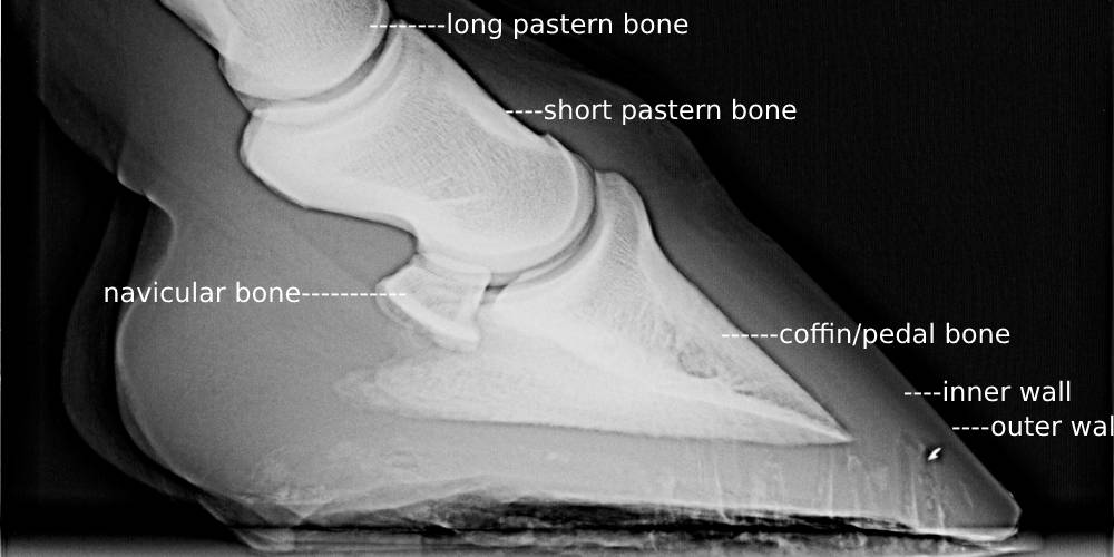
There are many different parts of the hoof both internally and externally. After writing my laminitis article and the article on parts of the hoof, I decided that showing the skeletal structure of the hoof could be beneficial.
List Of The Internal Anatomy Of The Hoof
There are several bones that make up the inside of a horse’s hoof. These are:
- Coffin/Pedal Bone
- Navicular/Sesamoid Bone
- Short Pastern Bone
- Long Pastern Bone
There are also tendons, ligaments, and joints as well that make up the inside of the hoof. These are:
- Deep Digital Flexor Tendon
- Coffin Joint
- Pastern Joint
- Superficial Flexor Tendon
- Sensitive Protective Lamina
Descriptions & Purposes For All The Internal Parts Of The Horse’s Hoof
Coffin/Pedal Bone
The Coffin bone, also known as the pedal bone, is the main bone found in the horse’s hoof. This bone is also what takes up much of the internal bone structure.
It is with this bone that issues can occur putting horses in life threatening situations. One of these is in the case of laminitis. Laminitis can cause the rotation of this bone to the point where the bone is pointing down and, in some cases, is even protruding out of the sole of the hoof.
(To read more on laminitis, click here!)
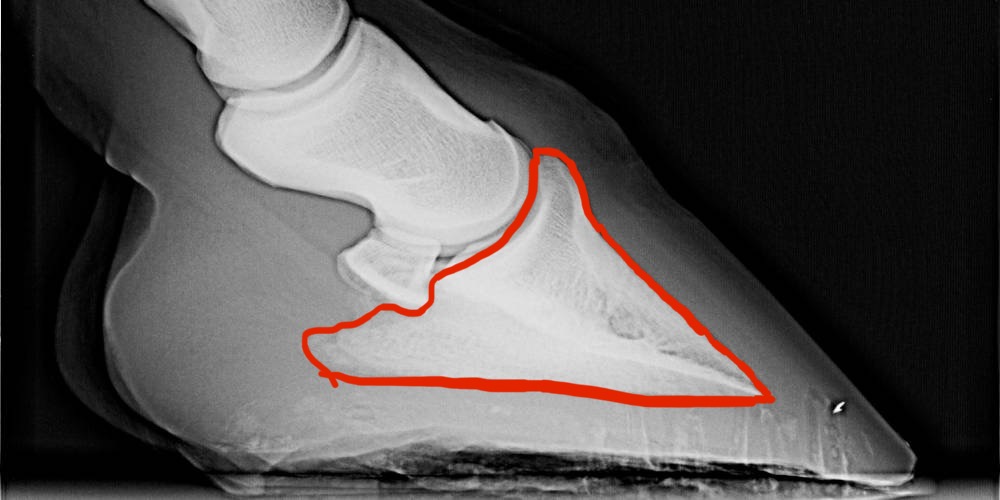
The coffin bone is in the center-most part of the hoof, basically the core of the hoof. It is, from the side, triangular in shape, but in overall appearance, it looks almost like a horseshoe crab.
In the following image, the coffin bone is outlined for easy identification:
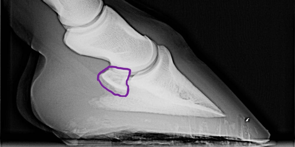
Navicular/Sesamoid Bone
The navicular bone, sometimes called the sesamoid bone, is found just behind the coffin joint. This small bone is held in place by a number of ligaments and it acts as a sort of fulcrum for the Deep Digital Flexor Tendon (DDFT).
This bone not only supports the DDFT, but it also acts as a form of support for the hoof. Sometimes, horses can suffer something known as Navicular Disease, also known as Navicular Syndrome. This syndrome or disease causes the degeneration of the Navicular bone causing the hoof to elongate and flatten along the ground due to the lack of support caused from the disappearance of the navicular bone. In many cases, Navicular Syndrome or Disease is the end of the road for horses. It is likely that horses with this illness will never be able to be ridden or worked again.
It was this bone that was shattered by the famous young racing filly known as Ruffian. In a race she was running against a Kentucky Derby winner, her sesamoid bones in her front right ankle shattered suddenly. Eight hours after this accident, Ruffian was euthanized from her condition.
In the image below, the Navicular bone is outlined for easier identification. For being so small, these bones are extremely important and based on the health of this bone, impact a horse’s entire life.
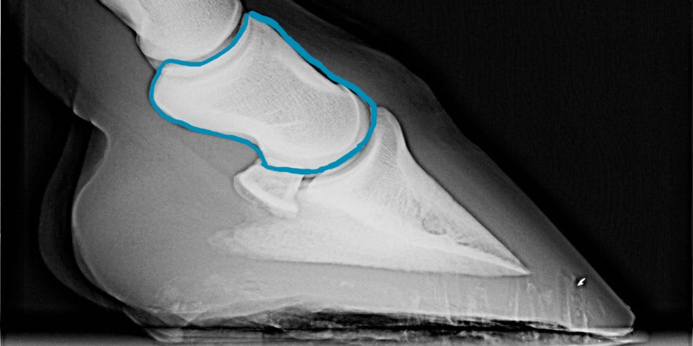
Short Pastern Bone
The short pastern is the bone that makes up the first part of the pastern. This bone connects to the coffin or pedal bone at the coffin joint while still contained in the hoof wall. It then connects to the long pastern bone at the pastern joint.
It is the short pastern bone that starts off the ‘leg bones.’ When looking at the parts of the horse’s leg, it is with this bone that the pastern of the horse begins. This bone, in an average sized horse, is roughly two inches in length.
It is mainly on this bone, specifically at the pastern joint, where horses can develop something known as ringbone. Ringbone is an issue seen mainly in older horses that causes bone growths on the bones and at the joints of the horse’s legs. This issue is similar to arthritis in the sense that bone-on-bone contact can be made. Often, if the ringbone is bad enough, the bony growths will grind on each other and cause immense pain and discomfort. In other cases, the ringbone can fuse a joint that stiffens the leg but results in no pain. If the ringbone is in a bad area of the leg, the horse may need injections to help the pain. Most horses with this condition can’t be ridden and may even need to be euthanized if their condition worsens over time.
In the following image, the short pastern bone is outlined:
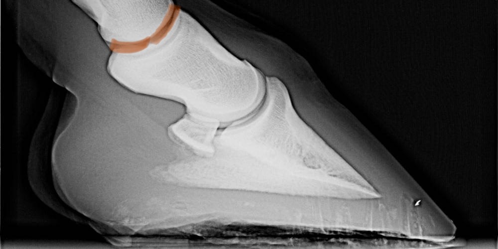
Long Pastern Bone
It is this bone that makes up most of the pastern of the horse before connecting to the fetlock joint. This bone is connected to the short pastern bone at something called the pastern joint.
This bone is preferably short rather than long. The reason for this is that this bone and the joints it is connected to act as the shock system for the horse’s body when it slams down onto the horse’s legs. If the long pastern bone is long, it is actually weaker as it is not as compact, and lameness issues involving the pastern are much more common.
In the image below, the bottom part of the long pastern bone is outlined to show where this bone connects of the short pastern bone at the pastern joint. This image should help you better understand the location of this bone:
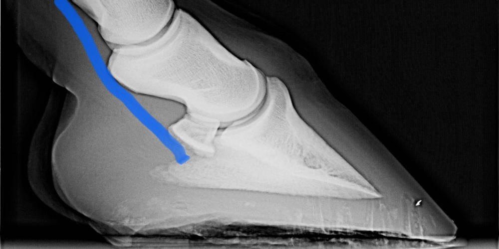
Deep Digital Flexor Tendon
Did you know that after the knee, there is actually no muscle in the horse’s legs at all??
All of the movement of the lower legs is caused by the use of tendons and ligaments such as the DDFT or the Deep Digital Flexor Tendon. This tendon is responsible for helping to lift the foot and is also involved in the fetlock joint movement as well.
There are a number of lameness issues surrounding the DDFT. One of the causes of the numerous health and conformation issues is caused from excessive tightness of this tendon. If the DDFT is too tight it can cause issues such as:
- Laminitis (to learn what this is click here!)
- Clubbed feet
- White Line Disease (briefly covered in my ‘parts of the hoof’ article. Click here to read it!)
- Navicular Inflammation (sometimes resulting in Navicular Syndrome or Navicular Disease)
In the image below, I drew the general location of this tendon in the hoof:
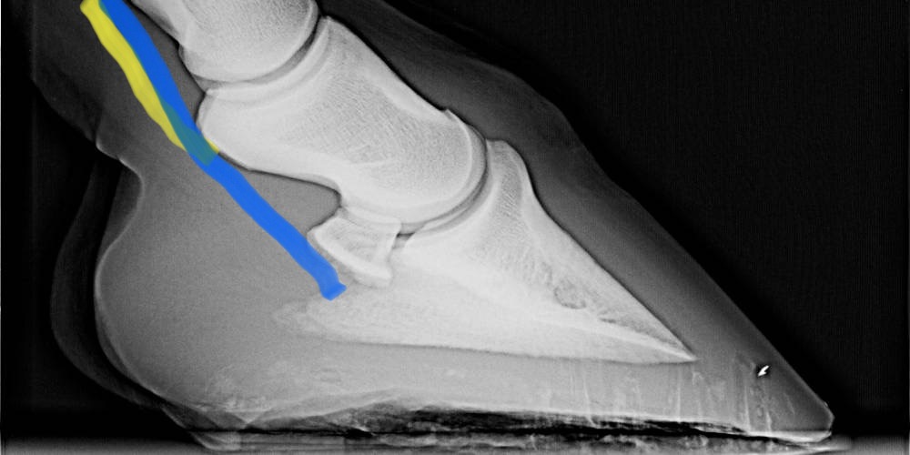
Superficial Flexor Tendon
Not only is there a Deep Digital Flexor Tendon, there is also a Superficial Flexor Tendon. This tendon is named superficial as it is closer to the surface of the skin that the DDFT is.
The Superficial Flexor Tendon is used to store energy. This tendon aids in the movement of the legs as does the DDFT, but because of the energy-storing aspect of this tendon, it is under significantly more pressure than the DDFT. Because of this, this tendon is much more prone to injury. Did you know that the tearing or damage of this tendon is actually the most common tendon injury seen in the horse world? The most common times for this tendon to become damaged is when a horse is jumping or racing. The constant high stress that is put on this tendon continuously in these sports is what causes such frequent injury.
The image below shows the location of two tendons. The blue line shows the DDFT as I talked about before while the yellow line shows the Superficial Flexor Tendon. I included them both in the same image to show the position of the tendons next to each other.
Coffin Joint
The coffin joint is a joint between the coffin bone and the short pastern bone in the hoof. This is a low motion joint found in the upper region of the hoof.
This joint can start to suffer pain and other issues if a horse begins to suffer Navicular Disease (also known as Navicular Syndrome). One of the treatments for Navicular Disease is actually to put an injection in the Coffin Joint.
This joint is also known to sometimes fuse if ringbone gets bad enough in a horse.
This joint is outlined in the image below:
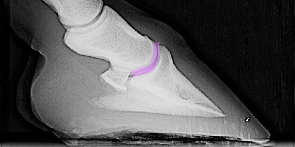
Pastern Joint
The pastern joint is a joint that is found between the long and short pastern bones. This joint is the most common location for the painful condition known as ring bone. This condition causes bony growths on certain bones and in certain joints causing excruciating levels of pain and lameness.
This joint is seen just above the top of the hoof just below the pastern itself.
The following image shows the location of the pastern joint:
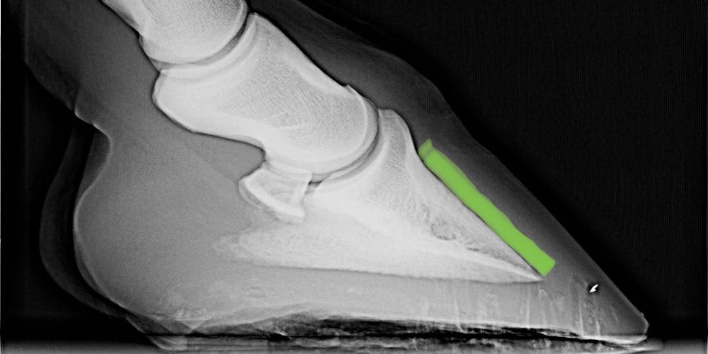
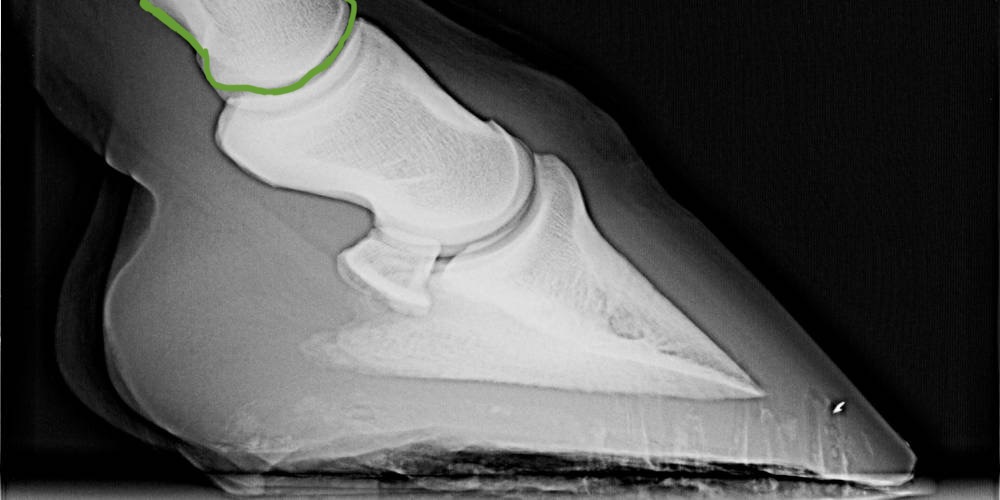
Sensitive Protective Lamina
The lamina of the hoof is a protective layer that falls between the front of the coffin bone and the hoof wall. This layer is just there for pressure absorption and protection, but it can also cause a number of problems.
If the protective lamina gets swollen, in the case of founder or laminitis, it can cause the coffin bone to actually rotate and protrude out of the bottom of the hoof. If this happens, the horse must be euthanized.
In the image below, the green marking indicates where the protective lamina is located in the hoof: